


Anna Grochot-Przeczek is an associate professor in theDepartment of Medical Biotechnology, Faculty of Biochemistry, Biophysics, and Biotechnology, Jagiellonian University in Krakow, Poland. She studies the molecular mechanisms that regulate the function of endothelial cells and blood vessels with a focus on NRF2/KEAP1 pathway, ageing and protein S-nitrosation. Currently, she investigates the importance of NRF2/KEAP1 imbalance and loss of proteostasis in the function of blood vessels.

Manuela G. Lopez is MD PhD and full professor of Pharmacology at the Department of Pharmacology in the School of Medicine, Universidad Autónoma de Madrid (UAM), Spain. Currently, she heads the Institute Teofilo Hernando for drug discovery (http://www.ifth.es/ ) that belongs to UAM. Her group, “NeuroprotectionLab” (http://neurodiscovery-ndd.com/gt1), has particular interest in the identification of new potential therapeutic targets to develop innovative and disease modifying therapies for neurodegenerative diseases, with special focus in modulating neuroinflammation (microglia-astrocyte interaction), oxidative stress and autophagy. Within the field of NRF2, she has contributed to the understanding of NRF2 in pain, depression, stroke and neurodegenerative diseases, together with the development of different NRF2 multitarget drugs in collaboration with medicinal chemists. Currently, she is coordinating a drug development project to identify Keap1-NRF2 inhibitors with potential use in Alzheimer’s disease.
NRF2 and Tumor Immunity
Anna-Liisa Levonen¹
¹A.I.Virtanen Institute for Molecular Sciences, Faculty of Health Sciences, University of Eastern Finland, Kuopio, Finland
e-mail: anna-liisa.levonen@uef.fi
Due to increase in proliferation and cellular metabolism, malignant cells are prone to oxidative stress, which renders pathways with antioxidant effects under positive selection. The principal transcriptional regulator of cellular redox homeostasis, NRF2 (NFE2L2 gene), is frequently hyperactive in malignant disease through e.g. somatic mutations in KEAP1 or NFE2L2. Along with its antioxidant effects, sustained NRF2 activity promotes cancer cell survival via other mechanisms, such as metabolic reprogramming and enhanced phase II enzyme expression causing resistance to radio- and chemotherapy. There is also mounting evidence that aberrant activation of NRF2 in cancer cells impacts local antitumor immune responses and is associated with impaired response to immunotherapy, yet the mechanisms remain unclear. Conversely, how NRF2 in tumor microenvironment affects cancer progression is ill understood, yet NRF2 has an acknowledged role in the regulation of inflammation and immunity. Herein, to address the effect of cancer cell-intrinsic NRF2 activity on tumor immunity, we developed an NRF2 activity metric and used it to conduct a pan-cancer analysis of oncogenic NRF2 signaling. To examine the contribution of NRF2 status within the host, a mouse model was employed whereby primary lung cancer cells expressing mutant Kras in combination with Tp53 loss were transferred to either wild type, Nrf2-deficient or Keap1 hypomorphic mice with moderately elevated NRF2 activity in order to address the role of stromal cell NRF2 on metastatic lung cancer growth. Spatial transcriptomics and proteomics techniques were used to study cell type-specific gene and protein expression. In the cancer-wide analysis of publically available datasets, we identified an immunoevasive phenotype where high NRF2 activity is associated with low interferon-gamma, HLA-I expression and T cell and macrophage infiltration in squamous malignancies of the lung, head and neck area, cervix and esophagus. In studies addressing the role of host Nrf2 on metastatic lung cancer growth, it was found that the Nrf2-deficient mice developed significantly fewer metastasis in the lung and had better survival than either wild type or Keap1 hypomorphic mice, with differences in cellular composition and gene expression of tumors between genotypes. To conclude, both cancer cell and stromal NRF2 status affect cancer progression, with important implications for the development of drugs modifying NRF2 activity.

Anna-Liisa Levonen is currently Professor and Vice Dean of Research at the University of Eastern Finland, Faculty of Health Sciences. She is also Associate Editor of Redox Biology. Her research program focuses on the gene regulatory mechanisms activated by oxidative and electrophilic stress, particularly via the redox-activated transcription factor NRF2. She has studied the mechanism of activation of the KEAP1-NRF2 pathway and its role in disease, particularly cancer and cardiometabolic diseases, and has published highly cited original and review articles on the topic.
Harnessing NRF2: Bridging In Vitro and In Vivo Insights for Age-Related Macular Degeneration Therapy
Ana S Falcão, Margarida Pedro, Lauida de Lemos, Shuvajit Rakshit, Pedro Antas, Sandra Tenreiro and Miguel C. Seabra
iNOVA4Health, NOVA Medical School|Faculdade de Ciências Médicas, NMS|FCM, Universidade Nova de Lisboa; 1169-056 Lisboa, Portugal.
email: stenreiro@nms.unl.pt
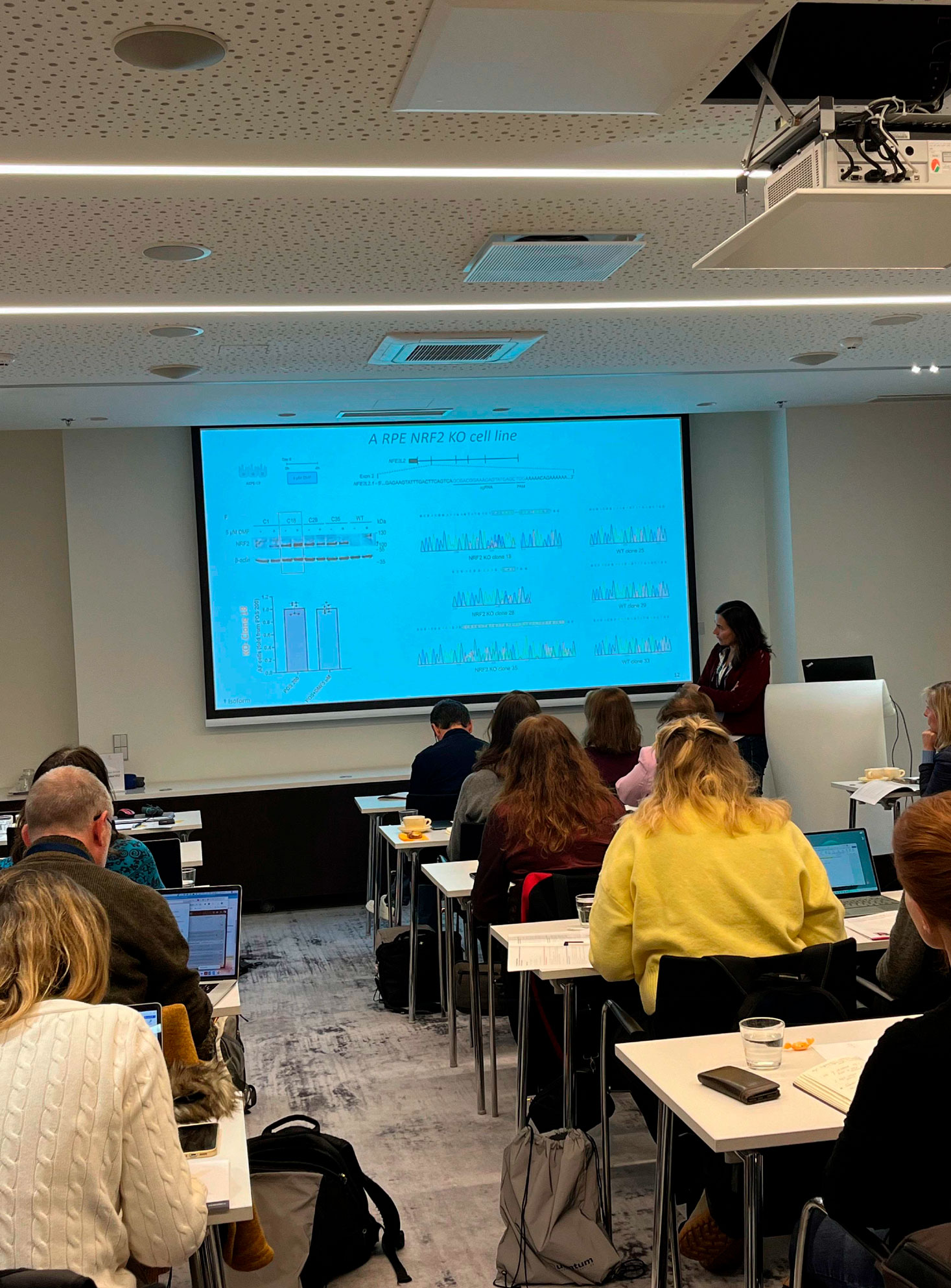
Age-related macular degeneration (AMD) is the most common blinding disease in the western world and is currently incurable. The primary cause of pathology appears to be retinal pigment epithelium (RPE) thinning, accumulation of autofluorescence (lipofuscin) and depigmentation, leading to atrophy and accumulation of extracellular deposits called drusen between the RPE and the choroid. The RPE is responsible for the daily digestion of photoreceptor outer segments (POS), which imposes a heavy continuous burden on the lysosomal network. Among the cell damaging events is photodamage of lipofuscin which may induce oxidative stress (OS). In fact, the retina is especially prone to OS due to high oxygen pressure and exposure to UV and blue light promoting reactive oxygen species generation. We have developed a model system that recapitulates some AMD features in vitro; feeding RPE monolayers in culture with POS leads to accumulation of autofluorescent granules similar to lipofuscin in vivo, resembling RPE stress and disease. Indeed, the activity of NRF2 in the RPE of aged mice has been found to be impaired, indicating that the aging RPE has a limited capacity to respond to perturbations in OS levels, and suggesting that an impaired anti-OS response by aging RPE after an oxidative stimulus could induce RPE dysfunction that may contribute to the development of AMD. Our preliminary results showed that overexpression of the transcriptional regulator NRF2, as well as treatment with NRF2 activators, can prevent the formation and/or resolve POS-derived autofluorescent granules, suggesting that this pathway and the proteins it controls are important for autofluorescent granule formation. We recently developed a stable NRF2 KO using CRISPR/Cas9 technology in an RPE cell line where we are testing the specificity and mechanisms of action of newly identified and repurposed NRF2 activators. In parallel, we are starting in vivo studies using an inducible mice model of AMD, to validate the data obtained in vitro. Our most recent results using these AMD models will be presented and discussed with the ultimate goal of validating Nrf2 as a new therapeutic target for AMD. Project funded by “La Caixa Foundation” (NASCENT HR22-00569)

Sandra Tenreiro heads the Degeneration and Ageing Lab at NOVA Medical School, Universidade Nova de Lisboa, Portugal, since April 2022. Her research is primarily focused on unraveling the molecular mechanisms behind retinal degeneration within the framework of aging-related diseases, utilizing retinal organoids. Sandra holds a BSc in Biochemistry, an MSc in Cell Biology, and a PhD in Biotechnology. She currently leads the Retina & Vision thematic research line in her institute, which encompasses seven research groups comprising 28 PhDs and clinicians. Sandra has served as the Principal Investigator (PI) and Co-PI in several research projects and has been extensively involved in advanced training, overseeing post-doctoral researchers, PhD candidates, and MSc students. Additionally, she collaborates with various PhD and MSc programs at both Universidade Nova de Lisboa and Coimbra University. Overall, her scientific career is exemplified by 57 peer-reviewed publications, with an H-index of 31 as reported by Scopus.
The Hypothermia Puzzle: Decoding Its Molecular Effects
K.-L. Grassman¹, H. Vellama¹, F.M. Sirkel¹, T. Jagomäe¹, L. Tarve¹, T. Visnapuu¹, R. Reimets¹, C.A. Hundahl¹, E. Vasar¹, H. Luuk¹, K.-L. Eskla1
¹University of Tartu, Institute of Biomedicine and Translational Medicine, Department of Physiology, Tartu, Estonia
email: kattri-liis.eskla@ut.ee
Ischemia reperfusion injury remains a significant challenge in various clinical settings, necessitating effective therapeutic interventions. Hypothermia has emerged as a promising strategy for mitigating cellular damage associated with oxygen deprivation. Our study investigates the molecular mechanisms underlying hypothermia-mediated protection using an in vitro model system. We demonstrate that hypothermia exerts a dual effect on cellular metabolism, simultaneously suppressing metabolic activity while enhancing stress tolerance. Specifically, mild hypothermia at 32°C emerges as the optimal temperature for balancing these opposing effects. Mechanistically, hypothermia attenuates hypoxia-induced metabolic disturbances, including alterations in lactate:pyruvate ratios and gene expressions associated with reductive stress. Notably, our research reveals a previously unrecognized role of hypothermia in modulating gene expression, with over 3000 genes responding to cooling, surpassing the response to hypoxia. Intriguingly, hypothermia-induced activation of hypoxia response element (HRE) transcriptional activity is dependent on HIF1B, independent of HIF1A or HIF2A. At 32°C, hypothermia activates Nrf2, promoting thioredoxin and glutathione-related gene expression and elevating glutathione levels. Furthermore, pre-conditioning with hypothermia enhances cellular resilience against oxidative stress. Current evidence highlighting potentially therapeutic molecular mechanisms can be translated to in vivo models, for further evaluation of therapeutic efficacy. Understanding the interplay between hypothermia, metabolic regulation, and stress response pathways holds promise for developing

Kattri-Liis Eskla In 2019, she earned her PhD in neuroscience from the University of Tartu (Estonia), discovering hypothermia’s profound impact on the antioxidant system. Her journey took her to Emory University School of Medicine (USA), where she explored novel treatments for cardiovascular diseases involving hydrogen sulfide and therapeutic lymphangiogenesis. Transitioning to the University of Birmingham (UK) in 2021, she embarked on collaborative research into hypoxic biology. Currently, her work challenges conventional notions about hypothermia, suggesting that its therapeutic effects are not solely due to metabolic depression. She is dedicated to unraveling the mechanisms behind hypothermia and reductive stress, aiming to transform the way we approach treating hypoxic/ischemic conditions (stroke, heart attack, organ transplantation, HIE) and disorders associated with electron transport chain dysfunction and reactive oxygen species formation.
Activation of NRF2 pathway protects against lipotoxicity in Protein tyrosine phosphatase 1B-deficient liver progenitor cells under steatohepatitis environment
M. Pilar Valdecantos1,2, Silvia Calero-Pérez1,2, Inés Barahona1,2, Alma Astudillo, Jesús Balsinde2,3, Ángela M. Valverde 1,2.
1IIBm Alberto Sols (CSIC-UAM), Madrid, Spain. 2CIBERdem (ISCIII), Spain. 3Instituto de Biología y Genética Molecular (CSIC-UVA), Valladolid, Spain.
email: avalverde@ib.uam.es
Activation of hepatic progenitor cells (PLCs) has been related to a regenerative response to repair liver damage during chronic liver diseases including non-alcoholic steatohepatitis (NASH). We have investigated the impact of palmitic acid (PA), a highly lipotoxic saturated fatty acid in LPC fate and the role of NRF2 in this context. PA induced apoptosis in LPCs in parallel to oxidative stress, mitochondrial damage and impaired autophagy. Analysis of nuclear and cytosolic NRF2 levels, expression of NRF2-target genes (Nqo1, Hmox1) and Heme oxygenase (HO-1) and Catalase protein levels, showed upregulated NRF2-mediated antioxidant defences and a rapid antioxidant response against lipotoxic stress in LPCs lacking protein tyrosine phosphatase 1 (PTP1B), encoded by the Ptpn1 gene. Interestingly, treatment of Ptpn1+/+ LPCs with PA plus sulforaphane recapitulated the protection against cell death found in Ptpn1-/- LPCs. We also evaluated the response of LPCs to the lipo-inflammatory environment of NASH. Treatment of LPCs with conditioned medium (CM) from macrophages stimulated with PA/LPS markedly reduced viability, effect attenuated in LPCs lacking Ptpn1. Interestingly, the CM collected from Ptpn1-/- LPCs previously exposed to PA/LPS-CM contained less proinflammatory mediators, suggesting their utilization to activate proinflammatory cascades that prime hepatocyte proliferation during liver regeneration. This was evidenced by the enhanced phosphorylation of STAT3, p65-NFκB, AKT and ERK together with IκBα degradation and p65-NFκB nuclear translocation in Ptpn1-/- LPCs. Moreover, under NASH-like conditions, basal/maximal respiration, ATP production and proton link were higher in Ptpn1-/- LPCs concurrently with an upregulation of antioxidant defences. In vivo studies in livers from mice fed a NASH-inducing diet revealed expansion of LPCs surrounded by inflammatory cells. Interestingly, mice transplanted with Ptpn1-/- LPCs showed higher expression levels of Krt19, a LPC marker, and less liver damage during NASH. Transcriptomic analysis revealed upregulation of hepatocyte and proliferation-related genes in Ptpn1-/- LPCs, reflecting a state of transitional liver progenitor cells that might offer therapeutic value to tackle NASH.

Ángela Martínez Valverde. My research career has is focused in molecular metabolism. From 2002 and as a Senior Scientist at Spanish National Research Council (CSIC) and CIBERdem (Spanish network in research on diabetes and related metabolic diseases) my laboratory investigates the mechanisms of insulin action in the liver and the molecular basis of metabolic-associated steatotic liver disease (MASLD, previously referred as NAFLD). We are interested in decipher the impact of insulin resistance, ER stress, autophagy, oxidative stress and inflammation in MASLD, as well as the interactome between liver cells in this context. Regarding therapeutics, we have unraveled the efficacy of a GLP1/Glucagon receptor co-agonist in improving liver regeneration during steatohepatitis. Our lab also studies the molecular basis of the metabolic alterations induced by chronic drug treatments, particularly the impact of antipsychotic medication on whole body metabolism and energy balance.
Disruption of Beta-TrCP/NRF2 Interaction. Relevance in Mouse Models of NASH
Raquel Fernández-Ginés1, José Antonio Encinar2, Ana I. Rojo1, Antonio Cuadrado1*
1Department of Biochemistry, Faculty of Medicine, Autonomous University of Madrid, Madrid, Spain;Autonomous University of Madrid. Centro de Investigación Biomédica en Red Sobre Enfermedades Neurodegenerativas (CIBERNED), ISCIII, Madrid, Spain. 2Institute of Research, Development and Innovation in Biotechnology of Elche (IDiBE) and Molecular and Cell Biology Institute (IBMC), Miguel Hernández University (UMH), 03202, Elche, Alicante, Spain.
email: antonio.cuadrado@uam.es
Transcription factor NRF2 is a master controller of several cellular homeostatic systems, such as antioxidant, anti-inflammatory mechanisms as well as metabolic regulation. These pleiotropic functions suggest that NRF2 might be a pharmacological target in several diseases. Much effort is being put on inhibiting the main NRF2 repressor, KEAP1, with either electrophilic small molecules or disrupters of the KEAP1/NRF2 interaction. The KEAP1 inhibitors omaveloxolone (Skyclarys) and several formulations of dimethyl fumarate (Tecfidera, Vumerity) have reached the market for the treatment of Friedrich ataxia, multiple sclerosis, and psoriasis. However, these KEAP1 inhibitors exert a very potent activation of NRF2 and maybe some of the unwanted side effects, such as liver damage, might be due to their strong induction of NRF2, highly beyond physiological levels. Our view is that acting on the weak inhibitor β-TrCP, leading to mild NRF2 activation, might provide long-term benefits during prolonged therapy in chronic diseases such as Metabolic dysfunction-Associated SteatoHepatitis (MASH). We are developing a new generation of mild NRF2 activators that selectively inhibits the interaction between β-TrCP and NRF2. Our lead compound, PHAR, upregulates NRF2-target genes such as Hmox1, Nqo1, Gclc, Gclm and Aox1, in a KEAP1-independent, but β-TrCP dependent manner, breaks the β-TrCP/NRF2 interaction, and inhibits the β-TrCP-mediated in vitro ubiquitilation of NRF2. Oral administration of PHAR elicits protective responses in the liver of two mouse models of non-alcoholic steatohepatitis. We suggest that mild NRF2 activation may provide a therapeutic strategy for diseases that present low-grade chronic inflammation, such as NASH.

Antonio Cuadrado is a full professor of Biochemistry and Molecular Biology at the Department of Biochemistry, Medical School, Autonomous University of Madrid. He obtained his PhD degree in Biology in 1985 and enjoyed several postdoctoral stays in the National Cancer Institute-NIH with the help of Fulbright and Fogarty fellowships. He established his independent laboratory as Professor of Biochemistry in 1997 with a main interest on the study of molecular mechanisms involved in initiation and progression of chronic diseases. For the past years his main lane of research has been the validation of transcription factor NRF2, master regulator of cellular homeostasis as a new therapeutic target in NASH. His current interest is the development of new NRF2-modulating drugs. Dr. Cuadrado has published over 160 primary and review articles, of which more than 80 are related to the role of NRF2 in physiological and pathological responses to disease.
Antioxidative Protection by Aripiprazole Does Not Induce Nrf2 Upregulation?
Irina Milisav1,2,*, Barbara Kramar1, Tinkara Pirc Marolt1, Dušan Šuput1
1Institute of Pathophysiology, Faculty of Medicine, University of Ljubljana, Slovenia 2Faculty of Health Sciences, University of Ljubljana, Slovenia
email: irina.milisav@mf.uni-lj.si
Aripiprazole and olanzapine are two second-generation antipsychotic drugs used to treat schizophrenia and bipolar disorders. Like other second-generation antipsychotics, their side effects include weight gain and higher blood sugar and lipids levels, frequently by olanzapine and much less by aripiprazole. Both drugs are metabolized in the liver. We examined their effect on the liver cells, primary hepatocytes and Fao hepatoma cell line. In contrast to clinical manifestations, aripiprazole has a more pronounced effect on the liver cells by increasing oxidative stress in the cells, with and without the addition of the oxidant. Increased oxidative stress in aripiprazole-treated cells boosts antioxidative responses, which does not seem to upregulate Nrf2 expression. Cells’ antioxidative protection is nevertheless an essential factor in cells’ tolerance to aripiprazole, a part of compensatory mechanisms that enable tolerance and safety of this drug. 1. Kramar B, Pirc Marolt T, Monsalve M, Šuput D, Milisav I. Antipsychotic Drug Aripiprazole Protects Liver Cells from Oxidative Stress. Int J Mol Sci. 2022 Jul 27;23(15):8292. doi: 10.3390/ijms23158292. 2. Pirc Marolt T, Kramar B, Bulc Rozman K, Šuput D, Milisav I. Aripiprazole reduces liver cell division. PLoS One. 2020 Oct 26;15(10):e0240754. doi: 10.1371/journal.pone.0240754. 3. Pirc Marolt T, Kramar B, Vovk A, Podgornik H, Šuput D, Milisav I. Therapeutic Dosage of Antipsychotic Drug Aripiprazole Induces Persistent Mitochondrial Hyperpolarisation, Moderate Oxidative Stress in Liver Cells, and Haemolysis. Antioxidants (Basel). 2023 Oct 30;12(11):1930. doi: 10.3390/antiox12111930.

Irina Milisav is a full professor of Biochemistry and Molecular Biology at University of Ljubljana, Slovenia, employed at Faculty of Medicine, Institute of Pathophysiology as a Research Associate and at Faculty of Health Sciences on a teaching post. She studies adaptive stress responses that boost cellular defences in liver cells (hepatocytes). Her group discovered an adaptive stress response of hepatocytes that prevents unnecessary apoptosis triggering when the stress-adapted hepatocytes are exposed to subsequent moderate stress (PACOS). ROS signalling mobilizes antioxidative defence and PACOS. Her group also investigates the effects of selected second-generation antipsychotics on the liver and the role of stress responses in drug-induced liver injury (DILI). She is a Grant Awarding Coordinator at COST CA20121 BenBedPhar Action.
NRF2 Activation by Electrophilic Metabolites
Albena T. Dinkova-Kostova¹ and Anna-Liisa Levonen²
¹Jacqui Wood Cancer Centre, Division of Cellular and Systems Medicine, School of Medicine, University of Dundee, Dundee, UK ²A.I. Virtanen Institute for Molecular Sciences, University of Eastern Finland, Kuopio, Finland
email: a.dinkovakostova@dundee.ac.uk
Under conditions of oxidative, inflammatory, and metabolic stress, cellular activity generates electrophilic metabolites, such as lipid oxidation and nitration products, and compounds derived from amino acid and central carbon metabolism. Many of these electrophiles activate transcription factor nuclear factor erythroid 2 p45-related factor 2 (NRF2), which in turn upregulates the expression of a network of genes encoding cytoprotective proteins that function to restore the cellular homeostasis. Most electrophiles activate NRF2 by chemically reacting with cysteine sensors within Kelch-like ECH associated protein 1 (KEAP1), the principal negative regulator of NRF2. The cyclopentenone prostaglandin 15-deoxy-Δ12,14-prostaglandin J2 (15d-PGJ2) and the unsaturated fatty acid derivative nitro-oleic acid modify C273 and C288 in KEAP1, whereas the reactive aldehyde 4-hydroxynonenal (4-HNE) modifies C151, C273 and C288. The Krebs cycle-related metabolites fumarate and itaconate alkylate C151. Other types of modifications of KEAP1 include S-lactoylation of C273 by glyceraldehyde 3-phosphate (Ga3P), N-succinylation of K131 by succinic anhydride, and formation of methylimidazole intermolecular crosslink between two KEAP1 monomers by methylglyoxal. Modified KEAP1 relays the initial signal to NRF2 and its transcriptional targets, the ultimate effectors that provide means for detoxification, adaptation and survival. Thus, these non-enzymatic covalent post-translational modifications in KEAP1 connect cellular metabolism with responses to stress and have functional consequences comparable to those caused by the more commonly known enzyme-catalyzed post-translational modifications.

Albena Dinkova-Kostova is a Professor of Chemical Biology at the University of Dundee School of Medicine (UK). She graduated in Biochemistry and Microbiology from Sofia University (Bulgaria) and obtained her PhD degree in Biochemistry and Biophysics from Washington State University (USA). She subsequently trained in Pharmacology at Johns Hopkins University School of Medicine (USA), where she continues to hold an Adjunct Professor position. She joined the University of Dundee in 2007 as a Research Councils UK Academic Fellow. Her group collaborates with basic scientists and clinicians, and with the pharmaceutical industry. In her research, at the interface of Chemical Biology and Medicine, she is committed to understanding how cells and organisms respond to oxidative, inflammatory, and metabolic stress, and is working towards development of strategies for protection against chronic disease. She was named among the top influential academics in Clarivate’s Highly Cited Researchers 2019, 2020, 2021, 2022 and 2023 lists.
siRNA-based Nanomedicines Targeting Nrf2 for Overcoming Cancer Drug Resistance
Stefania Pizzimenti1, Marie Angele Cucci1, Elisa Pettineo1, Debora Collotta1, Monica Argenziano2, Chiara Dianzani2, Roberta Cavalli2,
1Dep. Clinical and Biological Sciences, University of Turin, 10125 Turin, Italy 2Dep. Scienza e Tecnologia del Farmaco, University of Turin, 10125 Turin, Italy
email: stefania.pizzimenti@unito.it
It is well-known that nuclear factor E2-related factor-2 (Nrf2) plays an opposite role in cancer. At early stages, it has a tumor-suppressive action due to its ability to detoxify carcinogens, inhibit tumor growth, and protect cells from oxidative stress damage. However, at late stages, aberrant activation of Nrf2 is associated with poor prognosis. Indeed, Nrf2 activation inhibits apoptosis and enhances cell proliferation, as well as the self‐renewal capacity of cancer stem cells. Interestingly, Nrf2 participates in radioresistance, resistance to cytotoxic drugs, and targeted therapy. Indeed, its inhibition can overcome chemoresistance, thus making resistant tumor cells more sensitive to cytotoxic pro-oxidant drugs (i.e., cisplatin, doxorubicin), to radiotherapy or to targeted therapy drugs, such as dabrafenib and trametinib. This evidence strongly suggests that Nrf2 inhibition may be a promising therapeutic approach for advanced tumors. The use of a specific small interfering RNA (siRNA) targeting the Nrf2 gene to down-regulate its protein expression is an attractive strategy since it could entail greater specificity compared to pharmacological molecules. To make these molecules more stable and able to cross the lipophilic cell membranes, they can be loaded in nanovehicles (NV). Indeed, NV can protect siRNA from degradation during systemic circulation and transport siRNA to target cells, avoiding nonspecific delivery. Results obtained in our laboratories on siRNA-based nanomedicines (dendrimers and nanobubbles) targeting Nrf2 to overcome drug resistance to cisplatin, in melanoma and bladder cancer cells, and to targeted therapy (dabrafenib and trametinib) in melanoma will be presented.

Stefania Pizzimenti studied Biological Sciences at the University of Turin (Unito, Italy) (1992), holds a PhD in Experimental and Molecular Pathology (1997), and is a specialist in Clinical Pathology (2001). She is an Associate Professor of General Pathology at Unito. Her research topic focuses on oxidative stress in cancer. Initially, she investigated the role of lipid peroxidation products (e.g., 4-hydroxynonenal, HNE) in tumor progression. Nowadays, she is leading several research projects focusing on cellular signaling pathways (e.g., Nrf2, YAP) involved in oxidative stress and tumor progression, including chemoresistance, as well as the effect of nanoparticles carrying chemotherapeutic drugs or siRNA targeting Nrf2 in several tumor types.
Synthesis and Evaluation of Bile Acid-Derived NRF2 Activators Srđan Bjedov¹ ¹Department of Chemistry, Biochemistry and Environmental Protection, Faculty of Sciences, University of Novi Sad, Serbia Corresponding email: srdjan.bjedov@dh.uns.ac.rs
Nuclear factor erythroid 2-related factor 2 (NRF2) is a pivotal regulator of cellular defense mechanisms against oxidative stress and inflammation, making it a prime pharmacological target for various non-communicable diseases, including inflammatory, neurodegenerative, metabolic, and cancer conditions. The recent FDA approval of Omaveloxolone for Friedreich’s ataxia underscores the therapeutic potential of NRF2 activators. Steroidal compounds, by virtue of their biologically privileged structure, have shown significant interactions with the NRF2 pathway, exemplified by natural products and FDA-approved drugs such as Ursodeoxycholic acid, which modulate physiological functions through NRF2 engagement. In this study, we explore the synthesis of novel compounds based on the bile acid skeleton, specifically cholic acid, by incorporating enone and dienone moieties into the A ring of the steroidal structure, aiming to enhance NRF2 activation. The antioxidative properties of these synthesized compounds were evaluated through the induction of NRF2 target genes, including GCLC, GSR, GCLM, HMOX1, and NQO1, using quantitative PCR. Compounds SB140 and SB141 demonstrated substantial upregulation of these genes, suggesting potent NRF2 pathway activation. Their enhanced gene expression profiles imply a strong potential for therapeutic application in oxidative stress-related diseases. This presentation will detail the synthetic approach from cholic acid to the final compounds, their molecular interaction with NRF2, and the promising antioxidative capabilities observed. The findings support the continuing investigation into steroidal NRF2 activators and their potential for clinical application in managing a spectrum of NRF2-related diseases.

Srđan Bjedov is an assistant professor at the Department of Chemistry, Biochemistry and Environmental Protection, Faculty of Sciences, University of Novi Sad, Serbia. His research is focused on the synthetic and medicinal chemistry of mainly steroid compounds for the treatment of inflammatory, microbial, and cancer diseases.
Environmentally Friendly Synthesis of Carbon Dots for Cancer Therapy
Sofia Dominguez-Gil,¹ Yingru Zhou,1,2 Adalberto Camisasca,¹ Dorottya Krizsan,³ Susan J. Quinn,³ Alex J. Eustace,2 Silvia Giordani*¹
¹School of Chemical Sciences, Dublin City University, Glasnevin, Dublin, Ireland ²School of Biotechnology, Dublin City University, Glasnevin, Dublin, Ireland ³School of Chemistry, University College Dublin, Belfield, Dublin, Ireland
email: Silvia.Giordani@dcu.ie
Carbon dots, also known as carbon quantum dots or fluorescent carbon nanoparticles, are small-sized, spherical nanomaterials (below 10 nm) presenting amorphous carbon cores (mixed of sp2/sp3) with nanocrystalline regions of graphitic structure (sp2 carbon)1. They show unique properties such as higher aqueous solubility, robust chemical inertness, non-toxicity, and relatively low-cost manufacturing2. However, the efficient resistance to photobleaching and outstanding fluorescence are the most remarkable features compared to more conventional luminescent materials3. Here, we present a low-cost and environmentally friendly synthesis of carbon dots using natural resources like coffee grounds. The proposed synthetic route eliminates the use of highly toxic heavy metals and high energy-consuming reaction times, improving biocompatibility and benefiting the environment. These carbon dots were fully characterized by different techniques, showing small-size nanoparticles with tunable fluorescence. Biocompatibility studies on CAL-51 and SKOV-3 cell lines were also conducted showing good viability in both cellular lines. Furthermore, their surface functionalization makes them suitable for a wide range of applications in nanomedicine4 such as drug delivery shuttles, biosensors, or bioimaging5. (1) Szczepankowska, J.; Khachatryan, G.; Khachatryan, K.; Krystyjan, M. Carbon Dots – Types, Obtaining and Application in Biotechnology and Food Technology. Int J Mol Sci 2023, 24 (19). (2) Kurian, M.; Paul, A. Recent Trends in the Use of Green Sources for Carbon Dot Synthesis–A Short Review. Carbon Trends 2021, 3, 100032. (3) Cui, L.; Ren, X.; Sun, M.; Liu, H.; Xia, L. Carbon Dots: Synthesis, Properties and Applications. Nanomaterials 2021, 11 (12). (4) Behi, M.; Gholami, L.; Naficy, S.; Palomba, S.; Dehghani, F. Carbon Dots: A Novel Platform for Biomedical Applications. Nanoscale Adv. 2022, 4 (2), 353–376. (5) Bartkowski, M.; Zhou, Y.; Nabil Amin Mustafa, M.; Eustace, A. J.; Giordani, S. CARBON DOTS: Bioimaging and Anticancer Drug Delivery. Chemistry – A European Journal 2024, 11:e202303982.

Sofia Dominguez Gil completed her degree in Chemistry at the University of Valladolid (Spain). Afterwards, she carried out her Ph.D. and first postdoctoral research at the University of Montpellier under the supervision of Dr. Frédérique Cunin working on different types of nanoparticles for Rhabdomyosarcoma theranostic. Currently, she holds an Enterprise Partnership Postdoctoral Fellowship from the Irish Research Council to work at Dublin City University under the supervision of Prof. Silvia Giordani on the CAROTENE project. Her research project aims to develop new smart carbon-based nanomaterials capable of improving chemotherapeutic drug delivery.
Dimethyl Fumarate Treatment Prevented Social Stress-Induced Hypertension in Rats
Iveta Bernatova, Peter Balis, Silvia Liskova, Andrea Micurova, Michal Kluknavsky
Centre of Experimental Medicine, Slovak Academy of Sciences, Institute of Normal and Pathological Physiology, Bratislava, Slovakia,
email: Iveta.Bernatova@savba.sk
Dimethyl fumarate (DMF) is a known activator of nuclear factor erythroid 2-related factor 2 (NRF2, encoded by Nfe2l2 gene) function. Our ongoing studies indicate that long-term administration of DMF may have tissue-specific effects on NRF2-mediated mechanisms in experimental prehypertension. In this study, we investigated the effect of chronic oral administration of DMF on blood pressure, heart and vascular function of social stress-exposed adult male borderline hypertensive rats (BHRs). BHRs were offspring of spontaneously hypertensive dams and normotensive Wistar-Kyoto sires. They were exposed to crowding stress for six weeks with or without simultaneous DMF treatment (30 mg/kg/day, p.o.). Exposure to stress triggered elevations in blood pressure and reduced body weight gain in comparison to the control group. Concurrent treatment with DMF prevented the development of stress-induced hypertension. Exposure of rats to DMF or stress did not affect left heart ventricle weight when considered as the main effects of treatment in 2-way (stress x DMF) ANOVA analysis. The levels of conjugated dienes were reduced by both stress and DMF vs. control while gene expressions of Nfe2l2, Hmox1, Sod1 and Gpx4 were significantly elevated. In addition, there was a negative correlation between Nfe2l2 expression and levels of CD in the heart. However, simultaneous DMF plus stress exposure led to an increase in the expression of Tnfa, Il1b and Nos2. In the femoral artery, DMF did not alter endothelium-dependent nor endothelium-independent relaxations. However, DMF significantly reduced noradrenalin-induced contractions in stress-exposed rats. Thus, our data showed that DMF prevented the development of stress-induced hypertension via the mechanism associated with the modification of contractile functions in BHRs.

Iveta Bernatova is a senior scientist, serving as the Head of the Department of Experimental Hypertension and a Member of the Management Board at the Centre of Experimental Medicine, Slovak Academy of Sciences in Bratislava, Slovakia. She completed her undergraduate studies in Biochemistry in 1991, obtained her Ph.D. in Chemistry in 1997, and has held the title of Doctor of Science (D.Sc.) in Animal Physiology since 2009. Her research primarily focuses on the regulatory mechanisms of blood pressure in various experimental models of hypertension with particular attention to the role of nitric oxide and oxidative stress. She delves into the mechanisms underlying chronic social stress-induced hypertension and adaptation to stress. Her recent studies explore the effects of the NRF2 function activator DMF on the cardiovascular system in rats with varying genetic predispositions to hypertension. Iveta Bernatova has authored more than 120 peer-reviewed publications, which have garnered approximately 2000 citations. The current studies are supported by the projects VEGA No. 2/0157/21, MVTS-COST-CA20121 and APVV-22-0296.
Dimethyl Fumarate’s Influence on Tau-Induced Pyroptosis in Frontotemporal Dementi
Lilia A. Smith; Sara Kaminski-Santamaría; Aaron Abdelkader-Guillén; José Jiménez-Villegas, and Isabel Lastres-Becker,
Department of Biochemistry, School of Medicine, Universidad Autónoma de Madrid (UAM), Spain. Instituto de Investigación Sanitaria La Paz (IdiPaz), Instituto de Investigaciones Biomédicas “Alberto Sols” UAM-CSIC, Madrid, Spain.
email: ilbecker@ib.uam.es
Frontotemporal Dementia (FTD) is an early-onset, progressive neurodegenerative disorder affecting individuals under 65. FTD leads to profound changes in personality, cognition, and behavior/language due to frontal and temporal lobe degeneration, accompanied by hippocampal atrophy. Currently, there is no approved treatment to halt or slow down FTD progression. Recent research highlights the nuclear factor erythroid-derived 2-like 2 (NRF2) transcription factor as a crucial player in mitigating neurodegeneration and neuroinflammation. Notably, both FTD patient samples and a Tau22 transgenic model demonstrate activated pyroptosis, an inflammatory cell death pathway. This study explores the potential of the NRF2 inducer dimethyl fumarate (DMF) to reverse pyroptosis in our AAV-TAUP301L FTD model, offering a novel therapeutic avenue. In our established AAV-hTAUP301L mouse model, expressing human TAUP301L specifically in neurons via stereotaxic delivery, we aim to validate the therapeutic potential of dimethyl fumarate (DMF). This model, utilizing an adeno-associated viral vector (AAV9-hTAUP301L), induces tauopathy hallmarks, including gliosis and inflammation, mirroring human FTD within 21 days. Mice received oral DMF treatment (100 mg/kg) for 3 weeks. We assessed pyroptosis markers (NLRP3, ASC, GASDERMIN D, IL-1B, and IL-18rap) through quantitative PCR (qPCR) and immunofluorescence. Using bioinformatics analysis we determined whether these markers were NRF2-dependent genes. However, recent studies indicate that DMF is capable of inhibiting GASDERMIN D, modulating pyroptosis in an NRF2-independent manner. To determine the importance of this mechanism, we injected AAV9-TAUP301L into GASDERMINA D-deficient mice and analyzed both the cognitive impairment and the pyroptosis process. Our findings demonstrate that hippocampal TAUP301L overexpression triggers the pyroptosis pathway, with immunofluorescence revealing a preferential occurrence in microglia. Notably, DMF treatment effectively reverses pyroptosis, as evidenced by a significant reduction in all examined markers. DMF treatment effectively modulates the TAUP301L-induced pyroptosis process, unveiling an additional mechanism of action for this compound and highlighting its potential therapeutic application for individuals with FTD.

Isabel Lastres-Becker has more than 20 years of experience in the field of neurodegenerative diseases. Her work is focused on the molecular bases of neurodegeneration related to proteinopathy, neuroinflammation, and oxidative stress. She is the head of the laboratory of “New therapeutic strategies in neurodegenerative diseases: Parkinson’s disease (PD), tauopathies and amyotrophic lateral sclerosis (ALS)”. Since 2008 she has been interested in the implication of the transcription factor NRF2 in neurodegeneration, endorsed by 20 publications in relevant international journals. Her wide research background covers the most prevalent neurodegenerative diseases, looking for reliable markers of progression that can also serve as drug targets for modulating neurodegeneration such as NRF2. She is engaged in developing advanced new drugs and appropriate technology to establish treatments for those diseases, based on NRF2.
Three-Month Evaluation of a Sulforaphane-Generating Supplement Delivered through Drinking Water in Mice: Assessing Safety and Nrf2 Activation in Different Tissues
Panos G. ZIROS1, Georgios PSARIAS1, Sheng Huang1, Dionysios V. Chartoumpekis1, Massimo Bongiovanni2 and Gerasimos P. SYKIOTIS1
1Service of Endocrinology, Diabetology and Metabolism, Lausanne University Hospital and University of Lausanne, Lausanne, Switzerland 2Synlab Pathology, Lausanne, Switzerland
email: georgios.psarias@chuv.ch
Sulforaphane is one of the best studied Nrf2-activating compounds. In preclinical studies, it is usually administered either intravenously or via gavage. While these administration methods are efficacious in activating Nrf2 in target tissues, they have certain disadvantages: (i) they are invasive and thus not optimal from the perspective of the 3Rs (Replacement-Reduction- Refinement), notably regarding Refinement; (ii) their invasive nature may potentially affect certain molecular or behavioral readouts; (iii) they are labor-intensive for the personnel working with the experimental animals and require proper training to avoid injury to the animals; (iv) they do not recapitulate the preferred mode of administration of drugs to humans (i.e., oral intake). An oral sulforaphane-producing supplement (Avmacol®) in the form of a pill is on the market for human use. Each pill supplies broccoli seed extract that contains the sulforaphane precursor glucoraphanin as well as active myrosinase enzyme that converts glucoraphanin into sulforaphane in the small intestine. Studies in humans have supported that this supplement can effectively activate Nrf2 in vivo. The main objective of the present study was to test whether oral administration of the supplement to mice via the drinking water is safe and efficacious in activating Nrf2 in various target tissues. First, the supplement was dissolved in water and the capacity of the resulting solution to activate Nrf2 was tested by treating cells stably transfected with an ARE-luciferase construct. Dose-dependent Nrf2 activation was consistently observed. Maintaining the solution at room temperature for up to 7 days before treating the cells had no effect on its capacity to activate Nrf2 in vitro. Next, male and female adult mice were treated with the supplement, which was dissolved in their drinking water. The mice had continuous access to the supplement solution, which was their only source of drinking water. The solution was changed every 2-3 days, and the mice were treated for a total of 3 months. No adverse events or altered behavior were observed during this period. The mice were then sacrificed, and various tissues were harvested for molecular and histological analyses. A mild induction of Nrf2 activity was observed in the liver and other tissues, as reflected in an increase of Nqo1 mRNA expression levels. At the histological level, no signs of tissue damage were observed. These data demonstrate that administration of a commercially available sulforaphane-producing supplement to mice via the drinking water over 3 months is safe and efficacious in activating Nrf2. This method is then suitable for preclinical studies in the field of Nrf2-related research.

Georgios Psarias holds a Master of Pharmacy (MPharm) degree from the Department of Pharmacy at the University of Patras, Greece. He is presently engaged as a doctoral candidate within the Faculty of Biology and Medicine of the University of Laussane, in conjunction with the University Hospital of Lausanne (CHUV). His doctoral research centers on the functionality of the NFE2 family of transcription factors, specifically Nrf1 (NFE2-related factor 1) and Nrf2, investigating their pivotal roles in modulating cellular oxidative stress responses in thyroid tissues. While pursuing his master’s degree, Mr. Psarias participated in global contests held by MIT, centered around synthetic biology, especially the iGEM competition. Prior to his doctoral studies, he contributed to the Biology Department at the Hellenic Open University. His experience includes one-year research as an intern, during which he was integrally involved in a project regarding Epigenetics and their role in expression and Switch from Fetal to Adult Hemoglobin.
Transcriptomic and Epigenomic Profiling Reveals Altered NRF2 Responses to Diesel Emissions in Alzheimer’s Disease
Liudmila Saveleva1, Tereza Cervena2,4, Claudia Mengoni3, Michal Sima4, Zdenek Krejcik2, Kristyna Vrbov4, Jitka Sikorova2, Tosca O.E. de Crom5, Zuzana Šímová4, Mariia Ivanova1, Muhammad Ali Shahbaz1, Elina Penttilä6, Heikki Löppönen6, Anne M. Koivisto7,8, M. Arfan Ikram5, Pasi I Jalava9, Tarja Malm1, Sweelin Chew1, Michal Vojtisek-Lom2,01,12, Jan Topinka2, Rosalba Giugno3, Pavel Rössner4, Katja M Kanninen1
1A. I. Virtanen Institute for Molecular Sciences, University of Eastern Finland, Finland.2 Dept of Genetic Toxicology and Epigenetics, Institute of Experimental Medicine of the Czech Academy of Sciences, Czech Republic. 3Dept of Computer Science, University of Verona, Italy. 4Dept of Nanotoxicology and Molecular Epidemiology, Institute of Experimental Medicine of the Czech Academy of Sciences, Czech Republic. 5Dept of Epidemiology, Erasmus MC, University Medical Center, The Netherlands.6Dept of Otorhinolaryngology, University of Eastern Finland and Kuopio University Hospital, Finland. 7NeuroCentre, Kuopio University Hospital, Finland. 8Dept of Geriatrics and Dept of Neurosciences, Helsinki University Hospital, Finland. 9Dept of Environmental and Biological Sciences, University of Eastern Finland. 10Dept of Mechatronics and Computer Engineering, the Technical University of Liberec, Czech Republic. 11Faculty of Mechanical Engineering, Czech Technical University. Czech Republic.
email: Katja.Kanninen@uef.fi
Air pollution is a significant worldwide environmental risk factor for illness and premature death. Recent studies have correlated living close to major roads with risk of Alzheimer’s disease (AD). However, the mechanisms responsible for the link remain unclear. To investigate this, we employed an air-liquid-interface exposure system replicating real-world pollutant exposure and exposed human olfactory mucosa (OM) cells of healthy individuals and AD patients to diesel engine emissions (DE) and mRNA, miRNA expression and DNA methylation analyses were performed. DE exposure resulted in an almost four-fold higher responses in AD OM cells, indicating increased susceptibility to DE. Pathway analysis revealed significant activation of the NRF2 oxidative stress response and xenobiotic metabolism pathways in control cells. AD cells exhibited a differential response, including induction of heat-shock proteins. Different DNA methylation patterns were associated with gene expression changes, revealing new exposure targets. In vitro findings were validated by analysing data from the population-based Rotterdam Study, demonstrating that NRF2 signalling is activated and upregulated in individuals exposed to high levels of pollutants. Overall, this study provides new information on the adverse effects of DE in humans, identifies exposure biomarkers, and pinpoints key pathways involved activated by exposure. Moreover, the data suggest that AD individuals may face heightened risks due to impaired cellular defences, providing molecular insights into the air pollution-AD connection.

Katja Kanninen is a professor of Cellular Neurobiology at the A.I.Virtanen Institute for Molecular Sciences, University of Eastern Finland. She defended her thesis on NRF2 in Alzheimer’s disease and is now interested in understanding the interplay between environmental exposures such as air pollutants and NRF2. Her group works with in vivo models of neurodegeneration, and with both human and murine brain cells (primarily astrocytes and neurons), and human olfactory cells.
ACOD1 Modulation of Nrf2 Function in Treg in Sepsis
Michel Edwar Mickael1,2,*, Pavel Klimovich1, Atanas G. Atanasov1,3, Jarosław Olav Horbańczuk1, Piotr Religa4, Mariusz Sacharczuk1,5,* & Michał Ławińskii1,6,*
1Institute of Genetics and Animal Biotechnology, Postepu36a, 05-552, Garbatka, Poland 2Department of clinical immunology, PhiloMako research, Väpnaregatan 22 Linköping, Sweden ³LBI Digital Health and Patient Safety Medical University of Vienna Spitalgasse 23, Bauteil 86, 1090 Vienna, Austria 4Department of Medicine, Karolinska Institute, SE-171 77 Solna, Sweden. 5Department of Pharmacodynamics, Faculty of Pharmacy, Medical University of Warsaw, Banacha 1B, 02-091 Warsaw, Poland. 6Department of General Surgery, Gastroenterology and Oncology, Medical University of Warsaw, 02-091 Warsaw, Poland.
email: m.mickael@igbzpan.pl, michal-lawinski@wp.pl
Sepsis is a devastating disease that claims the lives of millions of people worldwide. The disease progresses from a simple reaction toward invading microbes to sequential organ failure leading to mortality. Patients who fall victim to sepsis pass through a phase of cytokine storms followed by a continuous cycle of proinflammatory responses led by Th17 and Th1, and an antiinflammatory response mediated by Treg cells. The reason behind Treg overactivation in sepsis is still unknown. We analyzed RNA-seq both on a bulk and a single cell level using cohorts of sepsis patients supported by animal KO studies to determine the main pathways controlling the Treg reaction in sepsis. We pinpointed the over-activation of metabolic pathways mediated by ACOD1 as the main cause behind Treg function in sepsis. Our analysis indicated that upstream regulation of ACOD1 is mediated by CD36 uptake of long fatty acid chains. Once activated, ACOD1 produces itaconate, which alkylates KEAP1, enabling NRF2 to activate various anti-inflammatory pathways. We hypothesize that targeting CD36-ACOD1-NRF2 through CD36 antagonists can prove valuable in breaking the cycle of organ failure in sepsis.

Michel-Edwar Mickael is an associate professor at the Institute of Genetics and Animal Biotechnology near Warsaw, Poland. His lab is interested in the Th17/Treg axis and its implications in autoimmune diseases. Michel learned several experimental and computational techniques while doing Ph.D. and postdoc training at Durham University, Karolinska Institutet, Victor Babes Institute, UKE (Germany), and UAB (Alabama).
The Heme Oxygenase-1/Carbon Monoxide System Modulates Bioenergetic Metabolism in Microglia
Roberta Foresti¹, Hiroaki Kitagishi², Roberto Motterlini¹
1Faculty of Health, University Paris-Est Créteil, INSERM, Créteil, France ²Department of Molecular Chemistry and Biochemistry, Faculty of Science and Engineering, Doshisha University, Kyotanabe, Kyoto 610-0321, Japan
email: roberta.foresti@inserm.fr
Microglia are important cells that exert immunological functions in the brain and acquire pro- and anti-inflammatory profiles during pathological stresses. It is has been established that cellular metabolism is directly implicated in microglia activation, favoring glycolysis over oxidative phosphorylation during a pro-inflammatopry state. Heme oxygenase-1 (HO-1) and its product carbon monoxide (CO) exhibit anti-inflammatory actions, which may be the result of their direct contribution on energetic metabolism. We will present experimental evidence supporting a role for HO-1/CO in the modulation of mitochondrial function and a regulatory role of CO against lipopolysaccharide (LPS)-induced mitochondrial dysfunction. Microglia cells exposed to hemin, and inducer of HO-1 and substrate for heme oxygenase activity, increased cellular respiration, while siRNA against HO-1 expression suppressed this effect. Furthermore, R8-HemoCD1, a mitochondrial-directed probe that scavenges CO with extremely high affinity, reduced oxygen consumption in a concentration-dependent manner in untreated cells and prevented the increase in respiration induced by hemin. Interestingly, intracellular ATP levels were also modulated under these conditions, indicating that endogenously-produced CO exerts signaling activities in mitochondria with functional consequences on energy levels. Microglia cells exposed to exogenous CO liberated by the a CO-releasing molecule (CORM-401) exhibited an opposite effect on mitochondrial respiration depending on the concentration used, as low CORM-401 increased oxygen consumption rate while higher amounts inhibited respiration. CORM-401 counteracted LPS-induced depression of microglia respiration and reduction in ATP levels. The early expression of inflammatory markers was also altered, suggesting that metabolic changes induced by CO are associated with control of inflammation. Mechanistic studies at molecular levels indicated a specific effect of CO on respiratory complex activities after challenge with LPS. Altogether, these data support a role for CO in controlling mitochondrial function under normal and inflammatory conditions.

Roberta Foresti (PhD) is Professor of Biochemistry at the Faculty of Health at the University Paris Est Créteil and working as a scientist at the Mondor Institute of Biomedical Research (IMRB). Dr Foresti teaches different subjects, such as human nutrition, energetic metabolism and oxidative stress, to bachelor and master students. She has dedicated more than two decades to investigating the protective action of the heme oxygenase-1 (HO-1) pathway in the cardiovascular system and inflammation, additionally focusing on drug discovery approaches targeting Nrf2/HO-1. She contributed to demonstrate an important role for the HO-1-derived products bilirubin and carbon monoxide (CO) in the protection of cardiovascular function during stress and is currently interested in the study of CO in obesity and metabolism. Dr Foresti is responsible of International Relations at IMRB, organizing a series of activities for the international recognition of our institute, including a summer school on environmental aggressions and health/chronic disease.
Hypothalamic Regulation of Energy Homeostasis and Obesity
Yavuz Yavuz1 & Bayram Yilmaz1,2
¹Yeditepe University, Faculty of Medicine, Department of Physiology, Istanbul, Türkiye ²Izmir Biomedicine and Genome Center, Izmir, Türkiye
email: bayram2353@yahoo.com
Obesity is an increasing global and major public health problem. The fundamental cause of obesity is the energy imbalance between calorie intake and expenditure. However, genetics, individual, social and environmental factors also play role in the etiology of this complex metabolic disorder. The hypothalamic arcuate nucleus (ARC) Agouti-Related Peptide (AgRP) and Proopiomelanocortin (POMC) neurons play important roles in hunger and satiety, respectively (Sayar-Atasoy et al, 2024). Our recent findings have shown the importance of Tyrosine hydroxylase (TH) neurons in the hypothalamic feeding pathways (Aklan et al, 2020). These three circuits process and maintain energy balance. Signaling between the brain and peripheral factors is usually impaired in obesity and metabolic syndrome. Recent discovery of opto/chemogenetic techniques have allowed neuroscientists to better elucidate the hypothalamic circuits controlling energy metabolism by selective excitation or inhibition of specific neuron groups. Use of viral vectors and transgenic mice technology are very useful tools in examining AgRP, POMC and TH projections and circuitry in the brain. In this talk, utilization of electrophysiology (including patch-clamp technique), opto/chemogenetic and fluorescent microscopy methods in neuroendocrine research will be reviewed. Acknowledgement: This study was supported by TÜBİTAK (Projects #118S245 and #122S382). References: Aklan I, Sayar Atasoy N, Yavuz Y, Ates T, Coban I, Koksalar F, Filiz G, Topcu IC, Oncul M, Dilsiz P, Cebecioglu U, Alp MI, Yilmaz B, Davis DR, Hajdukiewicz K, Saito K, Konopka W, Cui H, Atasoy D. (2020). NTS catecholamine neurons mediate hypoglycemic hunger via medial hypothalamic feeding pathways. Cell Metabolism, 31: 313-326.e5. Sayar-Atasoy N, Aklan I, Yavuz Y, Laule C, Kim H, Rysted J, Alp MI, Davis D, Yilmaz B, Atasoy D. (2024). AgRP neurons encode circadian feeding time. Nature Neuroscience, 27: 102-115.

Bayram Yilmaz is a Professor of Medical Physiology at Yeditepe University in Istanbul, Türkiye. He has a Doctor of Veterinary Medicine degree from Firat University and PhD in Medical Physiology from University of Glasgow. He worked as a Research Assistant Professor at the State University of New York at Albany, and Visiting Scientist at the King’s College London and at the University of Oxford. He is an author of seven book chapters and 152 refereed articles (h-index 32, >2900 citations). His research interests include neuro-endocrine regulation of energy homeostasis and reproductive hormones in experimental models. He also works on presence of endocrine disrupting chemicals in human breast milk and environmental samples using in vitro and in vivo test models. He is currently the Director of Izmir Biomedicine and Genome Center in Izmir, Türkiye.
Sustained Nrf2 Overexpression-Induced Metabolic Deregulation Can Be Attenuated by Modulating Insulin/Insulin-like Growth Factor Signaling
Sentiljana Gumeni, Maria Lamprou, Zoi Evangelakou, Maria S Manola, Ioannis P Trougakos Department of Cell Biology and Biophysics, Faculty of Biology, National and Kapodistrian University of Athens, Panepistimiopolis, 15784 Athens, Greece
email: itrougakos@biol.uoa.gr
The modulation of insulin/insulin-like growth factor signaling (IIS) is associated with altered nutritional and metabolic states. The Drosophila genome encodes eight insulin-like peptides, whose activity is regulated by a group of secreted factors, including Ecdysoneinducible gene L2 (ImpL2), which acts as a potent IIS inhibitor. We recently reported that cncC (cncC/Nrf2), the fly ortholog of Nrf2, is a positive transcriptional regulator of ImpL2, as part of a negative feedback loop aiming to suppress cncC/Nrf2 activity. This finding correlated with our observation that sustained cncC/Nrf2 overexpression/activation (cncCOE; a condition that signals organismal stress) deregulates IIS, causing hyperglycemia, the exhaustion of energy stores in flies’ tissues, and accelerated aging. We have extended these studies in Drosophila by assaying the functional implication of ImpL2 in cncCOE-mediated metabolic deregulation. We found that ImpL2 knockdown (KD) in cncCOE flies partially reactivated IIS, attenuated hyperglycemia and restored tissue energetics. Moreover, ImpL2 KD largely suppressed cncCOE-mediated premature aging. In support, pharmacological treatment of cncCOE flies with Metformin, a first-line medication for type 2 diabetes, restored (dose-dependently) IIS functionality and extended cncCOE flies’ longevity. These findings exemplify the effect of chronic stress in predisposition to diabetic phenotypes, indicating the potential prophylactic role of maintaining normal IIS functionality.

Ioannis Trougakos obtained his Ph.D. in Cellular-Developmental Biology from the National and Kapodistrian University of Athens (NKUA), Greece. He has worked as Research Scientist at EMBL, Germany, CBM “Severo Ochoa”, Spain and at NHRF, Athens, Greece; he was also research visitor at EMBL and at the Netherlands Cancer Institute. Dr. Trougakos was elected Research Lecturer at NHRF and currently he serves as Professor and Director of the “Cell Biology” lab at the Faculty of Biology, NKUA. He is the Head of the “Ageing and Age-Related Diseases” group (http://scholar.uoa.gr/itrougakos). Dr. Trougakos has published articles in high-ranking journals, chapters in international books; he is also co-inventor in several patents. His group is funded by private (GR, EU, USA) and public (GR, EU) entities; also, the group participates in contractual activities with the Industry.
Immunometabolic Responses Elicited by Sulforaphane in M1 (LPS) Polarizing Macrophages
Sheyda Bahiraii1,2, Martin Brenner1,2,3, Wolfram Weckwerth3 and Elke H Heiss1
1Department of Pharmaceutical Sciences, University of Vienna, Austria 2Vienna Doctoral School for Pharmaceutical, Nutritional and Sport Sciences, University of Vienna, Austria 3Vienna Metabolomics Center (VIME), University of Vienna, Austria
email: elke.heiss@univie.ac.at
Macrophage polarization towards the proinflammatory M1 (LPS) state involves expression of pro-inflammatory mediators and metabolic reprogramming. Distinct metabolic pathways not only serve as source of energy but also determine the functional state of immune cells (immunometabolism), suggesting deliberate metabolic rewiring as potential (adjuvant) strategy for immune modulation. This study shows that the Nrf2-activating and antinflammatory phytochemical sulforaphane (Sfn) influences metabolic shifts during M1 (LPS) polarization in murine macrophages in vitro. Compared to control M1 cells, Sfn administration leads to highly energetic cells showing both higher glycolytic and mitochondrial activity accompanied with an unbroken TCA cycle, elevated OXPHOS rate with fatty acids as preferred fuel, boosted fusion dynamics, lower membrane potential, and less superoxide production. Regarding the potential causative association between metabolic and anti-inflammatory features of Sfn in M1 (LPS) macrophages, Sfn reduces nuclear residence of dimeric pyruvate kinase (PKM)2, HIF-1α levels, Stat3 phosphorylation, and subsequently also IL-1β expression, while it preserves high levels of cytosolic PKM2 tetramer with high glycolytic enzyme activity. Moreover, sustained OXPHOS and TCA activity but not the concomitantly observed high dependency on fatty acids appeared necessary for Sfn to diminish expression of nos2, il1β, il6 and tnfα. M1 (LPS/Sfn) macrophages displayed lower nucleo/ cytosolic acetyl-CoA levels in association with lower histone acetylation at selected pro-inflammatory gene promoters. Overall, Sfn elicits distinct changes in cellular metabolism that may deserve increased appreciation as important feature of Sfn´s immunopharmacology, very likely also applicable to immune cells other than macrophages.

Elke Heiss is Associate Professor at the Department of Pharmaceutical Sciences at the University of Vienna, Austria. Prior to setting up her group in Vienna, she was trained as biochemist in Germany and the UK, did her PhD at the German Cancer Research Center followed by two postdoctoral stays in USA and Sweden. Her research activities focus on the elucidation of signal transduction events and in vitro pharmacology of small (natural) chemical entities, with particular focus on redox biology and the interface between cellular metabolism and function..
From Ayurveda to non-canonical ferroptosis: do phytochemicals have a future in precision oncology? Wim Vanden Berghe Cell Death Epigenetic Signaling Lab-IPPON, Department Biomedical Sciences, University Antwerp, Belgium Corresponding email: wim.vandenberghe@uantwerpen.be Website: https://www.kinases-epigenetics-ppes.com/
Disease relapse and therapy resistance remain key challenges in cancer treatment. Underlying (epi-)mutational events typically promote tumorigenesis and contribute to multi-drug therapy resistance. Although innovative biomarker “omic” strategies are instrumental to optimize personalized precision oncology treatments, dynamic epigenetic plasticity of (immunogenic) cell death pathways and cellular mosaicism of the tumor-immune ecosystem frequently cause therapy escape, which prevents complete recovery and ultimately triggers disease relapse. Interestingly, ferroptosis, a novel form of iron-dependent redox and lipid peroxidation-regulated cell death was recently identified as a novel appealing treatment strategy to overcome multidrug therapy resistance in different cancer types. New ferroptosis therapeutic strategies will be presented by redox bioactive phytomedicinal compounds, non-thermal plasma treatment and iron-binding heme proteins to overcome drug resistance. Finally, integrative systems biology (i.e. epigenome-transcriptome-kinome-proteome based) “kino2omic” approaches are being developed to predict ferroptosis therapy response which might advance the therapeutic applicability of ferroptotic compounds in future clinical cancer treatment precision medicine.

Prof. Wim Vanden Berghe obtained his PhD at the University of Ghent (UGent) in 1999 in the Faculty of Science Biochemistry- Biotechnology. After postdoctoral research at various research institutions (University of Montpellier-France, Stellenbosch- SouthAfrica and Oxford-UK), Wim Vanden Berghe was appointed professor of Epigenetics in 2009 at the cell death epigenetic signaling lab (CDES-IPPON) (UAntwerp/UGent, Belgium). With his current research, he is characterizing epigenetic plasticity of redox/kinase signaling pathways to modulate ferroptosis disease phenotypes with phytomedicinal or pharmacological compounds to improve treatment of cancer, CVD, obesity, NAFLD, autism spectrum disorders or neurodegeneration/development.